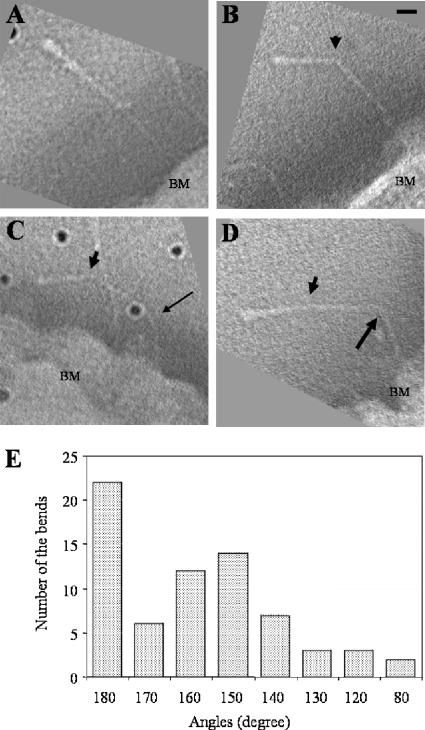
FIG. 2.
Projections through EmaA subvolumes extracted from the tomograms showing different morphologies of the EmaA appendages. Subvolumes have been rotated to visualize the EmaA appendage parallel to the imaging plane. (A) Straight EmaA appendage; (B) EmaA appendage with a bend at 29.4 nm (arrowhead) from the apical ending of the structure; (C) EmaA appendage with bends at 29.4 nm (arrowhead) and 65.6 nm (thin arrow); (D) EmaA appendage with bends at 29.4 nm (arrowhead) and 68.8 nm (thick arrow). BM, bacterial membrane. Bar, 10 nm. Dark points are gold particles. (E) Angular distribution of the bends at 29.4 nm.

