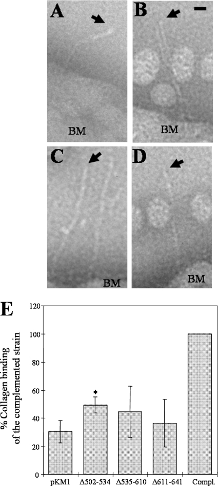
FIG. 3.
EmaA appendages windowed from electron micrographs of A. actinomycetemcomitans strains acquired from whole-mount preparations of the bacteria stained with 2% phosphotungstic acid (pH 7). (A) WT strain; (B) emaA mutant strain transformed with pKMΔ502-534; (C) emaA mutant strain transformed with pKMΔ535-610; (D) emaA mutant strain transformed with pKMΔ611-641. Arrows point to the apical end of the EmaA appendages. Spherical particles in the images are extracellular vesicles. BM, bacterial membrane. Bar, 10 nm. (E) Collagen binding activities of the emaA mutant strains in panels A to D and pKM9 (plasmid containing the full-length emaA gene and the upstream putative promoter region [Compl.]) as measured by enzyme-linked immunosorbent assay. The data are represented as the percentage of the binding of the full-length complemented strain (Compl.). *, statistically significantly different from the emaA mutant/pKM1 strain.

