FIG. 2.
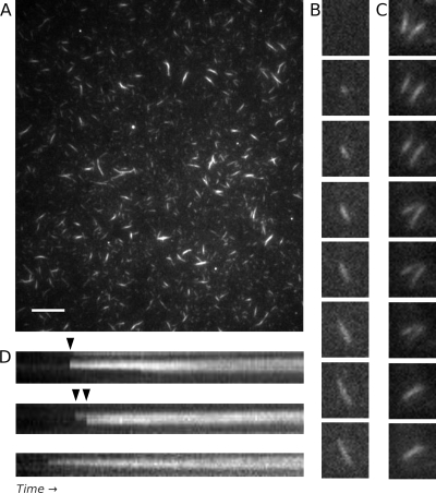
AlfA filament and bundle assembly observed by TIRF microscopy, with bundles showing no evidence of dynamic instability. (A) TIRF microscopy of a field containing Cy3- and biotin-labeled 3 μM AlfA 5 min after polymerization revealed stable filamentous structures with various fluorescence intensities. The variation in intensity is consistent with filament bundles. Scale bar, 10 μm. The conditions used were as follows: 20% Cy3-labeled AlfA and 5% biotin-labeled AlfA (total concentration, 3 μM) with 5 mM ATP in the buffer described in the legend to Fig. 1A. (B) Bundles appear to elongate bidirectionaly, but the polarity of the filaments (and the directionality of their elongation) cannot be determined. The time interval was 40 s. The conditions were the same as those described above, except that 0.5% bovine serum albumin was added. (C) Annealing between two bundles adhering to a coverslip. The time interval was 7 s. The conditions were the same as those described above for panel A. (D) Kymographs collected immediately after addition of nucleotide, showing that bundles can grow by lateral annealing events (arrowheads). The time scale was 2.5 min. The conditions were the same as those described above for panel A.

