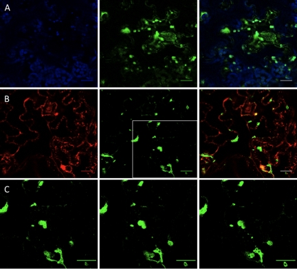
FIG. 2.
6K2GFP-tagged TuMV-induced vesicles. N. benthamiana cells expressing 6K2GFP-tagged TuMV-induced vesicles observed by confocal microscopy at 4 days after agroinfiltration. (A) Optical images (1-μm thick) showing 6K2GFP-tagged vesicles (green) with chloroplasts (in blue), with merge colors in the right panel. (B) Optical images (1-μm thick) showing 6K2GFP-tagged vesicles (green) and the Golgi marker fused to mCherry (red), with merge colors in the right panel. (C) Consecutive 1-μm thick single plane images (from left to right) of the depicted square in B that show GFP distribution within the vesicles. Scale bar, 10 μm.

