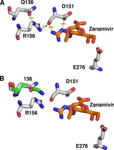
FIG. 3.
Modeling of resistance mutations based on the crystal structure of the H5N1-zanamivir complex. (A) Stick representation of wild-type H5N1 (white sticks) with zanamivir (orange sticks). (B) Mutation Q136K (green sticks) results in a loss of the hydrogen bond network. Figures were produced with the modeling software package PyMol (W. L. DeLano, DeLano Scientific, Paolo Alto, CA) using the sequence of the Protein Data Bank code 2HTQ protein (27) downloaded from the RCS Protein Data Bank.

