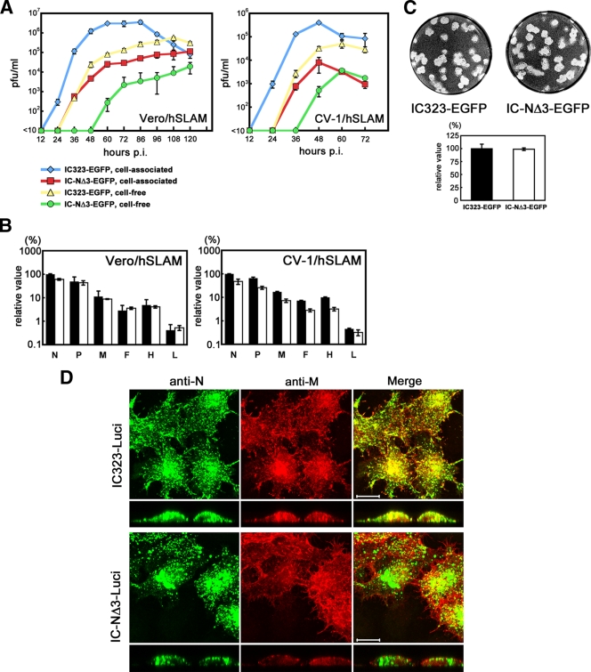
FIG. 4.
Analyses of recombinant MVs possessing carboxyl-terminal deletion of the N protein. (A) Growth kinetics. Vero/hSLAM and CV-1/hSLAM cells were infected with recombinant MVs (IC323-EGFP and IC-NΔ3-EGFP) at an MOI of 0.001. The infectious titers in culture medium (cell free) and cells (cell associated) were determined at various time points. (B) Quantification of viral mRNAs. Vero/hSLAM and CV-1/hSLAM cells were infected with IC323-EGFP (black bar) or IC-NΔ3-EGFP (white bar) at an MOI of 0.001 in the presence of a fusion-blocking peptide. At 24 h p.i., the levels of the N, P, M, F, H, and L mRNAs of MV in the infected cells were analyzed by reverse transcription-quantitative PCR. Data were normalized by the levels of β-actin mRNA and represent the means ± standard deviations of triplicate samples. The N mRNA level in IC323-EGFP-infected cells was set to 100%. (C) Plaque assays. Monolayers of CV-1/hSLAM cells on 12-well cluster plates were infected with 50 PFU of IC323-EGFP or IC-NΔ3-EGFP and overlaid with DMEM containing 7.5% FBS and 1% methylcellulose. At 5 days p.i., the cells were stained with 0.01% neutral red, and the sizes of the plaques were measured. The mean diameters ± standard deviations are shown in the bar graph. (D) HeLa/hSLAM cells were infected with IC323-Luci (upper panels) or IC-NΔ3-Luci (lower panels). At 24 h p.i., the intracellular distributions of the N and M proteins were analyzed by indirect immunofluorescence and confocal microscopy as described in the legend of Fig. 3A. Longitudinal sections are also shown at the bottom. Bar, 10 μm.

