FIG. 3.
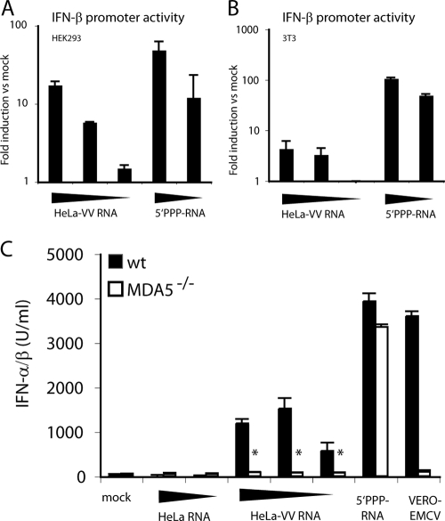
RNA from VV-infected cells stimulates MDA5. HEK293 (A) or 3T3 (B) cells were transfected with reporter constructs for IFN-β-luciferase and control plasmid pRL-TK and subsequently stimulated with HeLa-VV RNA (1, 0.2, and 0.04 μg) or control 5′ PPP-RNA (0.2 and 0.04 μg), respectively. Graphs show relative activation of the IFN-β promoter. Data are the average of duplicate measurements ± standard deviation. (C) MEFs of the indicated genotype were transfected with HeLa-RNA (1 and 0.2 μg), HeLa-VV RNA (1, 0.2, and 0.04 μg), Vero-EMCV RNA (0.2 μg), or with control 5′ PPP-RNA (0.2 μg). The graph shows the accumulation of IFN-α/β in supernatants after overnight incubation. *, P < 0.001 compared to wild-type (wt) MEFs. Data are average ± standard deviations of duplicate (A and B) or quadruplicate (C) measurements from one experiment repeated three times.

