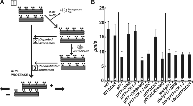
Figure 3.
Biochemical depletion of CK1 rescues microtubule sliding in isolated pf17 axonemes, and this rescue requires I1 dynein. (A) Experimental strategy to test the role of CK1 in microtubules. (B) ATP-induced microtubule sliding velocity was measured in isolated axonemes, CK1-depleted axonemes, or CK1-depleted axonemes reconstituted with purified rCK1 (Okagaki and Kamiya, 1986; Wirschell et al., 2009). The effect of DRB/CK1-7 and the phosphatase inhibitor microcystin-LR (MC) was also examined. The bars represent: (1) wild-type (WT) axonemes; (2) WT axonemes depleted of CK1 (note that there is no change in velocity); (3) pf17 axonemes (note the slow, baseline sliding velocity); (4) pf17 axonemes plus DRB; (5) pf17 axonemes plus CK1-7; (6) pf17 axonemes plus DRB and microcystin-LR; (7) pf17 axonemes plus CK1-7 and microcystin-LR; (8) pf17 axonemes depleted of CK1 (note the rescue of microtubule sliding); (9) pf17 axonemes depleted of CK1 plus microcystin-LR; (10) ida1pf17 axonemes; (11) ida1pf17 axonemes plus DRB; (12) ida1pf17 axonemes plus CK1-7; and (13) ida1pf17 axonemes depleted of CK1 (note the failure in rescue of sliding). Microtubule sliding velocity is expressed as µm/s, and means and standard deviations (error bars) were calculated from at least three independent experiments with a minimum sample size of 75 axonemes.

