Figure 2.
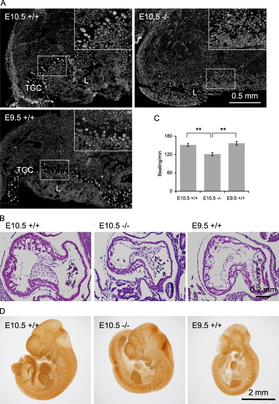
Characterization of Drp1−/− embryos. (A) DAPI-stained sections of placenta of age-matched E10.5 Drp1+/+ and Drp1−/− embryos and an E9.5 Drp1+/+ embryo, which was size matched to the E10.5 Drp1−/− embryos. Insets show enlarged images of boxed regions. Trophoblast giant cell (TGC) and labyrinth (L) layers are indicated. (B) H&E stains of sagittal sections of embryonic heart. (C) Beat rates in isolated embryonic cardiomyocytes were measured using differential interference contrast microscopy. (D) Whole-mount immunohistochemistry of embryos with anti-PECAM antibodies. **, P < 0.01 (n ≥ 39). Error bars indicate mean ± SEM.

