Figure 3.
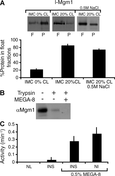
l-Mgm1 GTPase activity is inhibited when inserted into a membrane bilayer. (A) l-Mgm1 inserts into IMC liposomes. l-Mgm1 was reconstituted into IMC 0% (left) and IMC 20% CL (right) liposomes as described in Materials and methods and fractionated by floatation on sucrose gradients. A 0.5 M NaCl treatment was performed to remove uninserted l-Mgm1 before floatation (right). A representative SDS-PAGE and Western blot of equivalent amounts of the float (F) and pellet (P) fractions is shown. Quantification from three experiments is shown as the mean + SEM (error bars). (B) l-Mgm1 inserts in the correct orientation in IMC liposomes. Reconstituted l-Mgm1 liposomes were treated with trypsin in the presence and absence of MEGA-8 as described (see Materials and methods). (C) GTPase activity of l-Mgm1 was determined as described alone (left; NL, no lipids), after reconstitution into IMC 20% CL liposomes (left; INS), after reconstitution into IMC 20% CL liposomes and subsequent addition of 0.5% MEGA-8 (right; INS), and upon addition of detergent-solubilized l-Mgm1 to IMC 20% CL liposomes (right; NI). Data from three experiments are shown as the mean + SEM (error bars).

