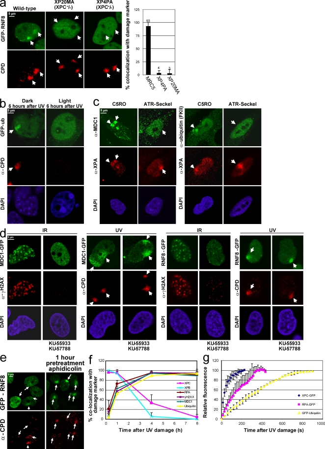
Figure 4.
NER intermediates trigger RNF8 accumulation in a DNA damage– and ATR-dependent manner. (a) RNF8-GFP stably expressing NER-proficient (MRC5) or NER-deficient (XPC-negative cell lines XP20MA and XP4PA) were locally UV exposed (60 J/m2). 2 h later, damaged cells were immunostained for CPDs. The percentage of cells in which RNF8-GFP colocalizes with CPD after LUD is plotted in the graph. RNF8 does not accumulate in NER-deficient cells. (b) Mouse embryonic fibroblasts expressing both CPD and 6-4PP photolysases were stably transfected with GFP-Ub. 1.5 h after local UV exposure (60 J/m2), cells were cultured either in the dark (left) or were photoreactivated with visible light for 2 h (right). 6 h after initial UV damage, cells were fixed and immunostained for CPDs. After UV lesion removal, GFP-Ub does not accumulate at LUD. (c) ATR hypomorphic human cells (Seckel) and C5RO cells with a wild-type ATR status were locally UV irradiated (25 J/m2) and immunostained after 2 h with antibodies recognizing MDC1 or conjugated Ub (FKII). XPA was used as a damage marker, indicating that MDC1 and Ub accumulate in an ATR-dependent manner. (d) Stably expressing MDC1-GFP (left) or RNF8-GFP (right) cells were incubated for 90 min with ATM and DNA-PK inhibitors (KU-55933 and KU-57788) or with an equal volume of DMSO. Cells were exposed to IR (10 Gy) or UV (60 J/m2), and after 2 h, they were immunostained for γH2AX or CPD. Although the addition of these inhibitors clearly inhibits the IR-induced foci formation of MDC1, MDC1 still accumulates after LUD. (e) Stable MRC5 RNF8-GFP–expressing cells were locally exposed to 20 J/m2 UV with or without a pretreatment for 1 h with 1 µg/ml aphidicolin. 1 h after UV damage, cells were fixed and immunostained for CPDs. A strong increase of RNF8 accumulation at the damaged DNA after aphidicolin pretreatment was observed. (f) The percentage of GFP-MDC1, XPB, RPA, γH2AX, GFP-Ub, and XPC-GFP colocalization with a damage marker (either CPD or XPA) at LUD (irradiated with 45 J/m2) is plotted for the different time points at 15 min, 1 h, 4 h, and 8 h after LUD. Although XPC and XPB are colocalizing in almost all cells 15 min after UV damage, the other factor colocalizes significantly with the used damage markers 1 h after LUD. (g) Cells stably expressing the XPC-GFP, GFP-Ub, and GFP-RPAp70 proteins were UV damaged using UV-C (266 nm) laser irradiation. GFP fluorescence intensities at the site of UV damage were measured by real time imaging until they reached a maximum. Assembly kinetic curves were derived from at least six cells for each protein. Relative fluorescence was normalized on 0 (before damage) and 100% (maximum level of accumulation). Arrows and arrowheads indicate local damage sites. Error bars indicate SEM.

