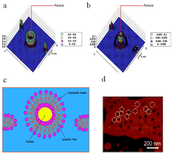
Figure 7.
Detailed proposed model of PAMAM dendrimer causing hole formation analogous to schematic models (ii) and (iii). (A) Simulated density map of the lipophilic part of the bilayer. (B) Simulated density map of the hydrophilic (headgroup) part of the bilayer. X-axis is normal to the bilayer plane. (C) Sketch demonstrating the formation of a nanoparticle-bilayer complex and corresponding breakage of the original membrane. (D) AFM pictures of lipid bilayers with holes made by charged PAMAM dendrimers. Particle radius Rp = 2.4 nm. Reprinted with permission from reference 10.

