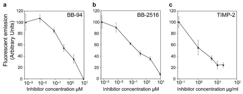
Fig. 4.

Quantitative analysis of the inhibition of cell surface metalloproteinase activity in human tumor cells. HT-1080 fibrosarcoma cells were seeded in a 96-well microtiter plate and treated for 60 min with 90 nM (5 μg/mL) LF/β-Lac and 26 nM (2 μg/mL) PrAg-L1 in the presence of increasing concentrations of the metalloprotease inhibitors BB-94 (a), BB-2516 (b), and TIMP-2 (c). Thereafter, 1.5 μM CCF2/AM was added to the cells for 60 min at room temperature. The CCF2/AM remaining in the medium was removed by washing, and the cells were incubated for 60 min at room temperature to allow for CCF2/AM hydrolysis. Fluorescence emission was recorded with a plate reader using 405 nm excitation and 460/25 nm bandpass for blue fluorescence and 535/25 nm bandpass for green fluorescence. The data are expressed as mean ± standard error of the mean of triplicate determinations. Reproduced in part from ref.12.
