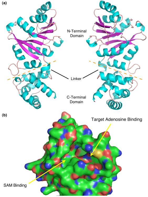
Fig. 2.
Structure of MjDim1. (a) Two views, rotated 180°, of the structure of MjDim1. α-helices are in cyan and β-strands are in magenta. The orange dashed line separates the two domains. The linker connecting the two domains is labeled, as are the domains. (b) A close up of the N-terminal domain highlighting the ligand binding pockets, as annotated.

