Figure 1.
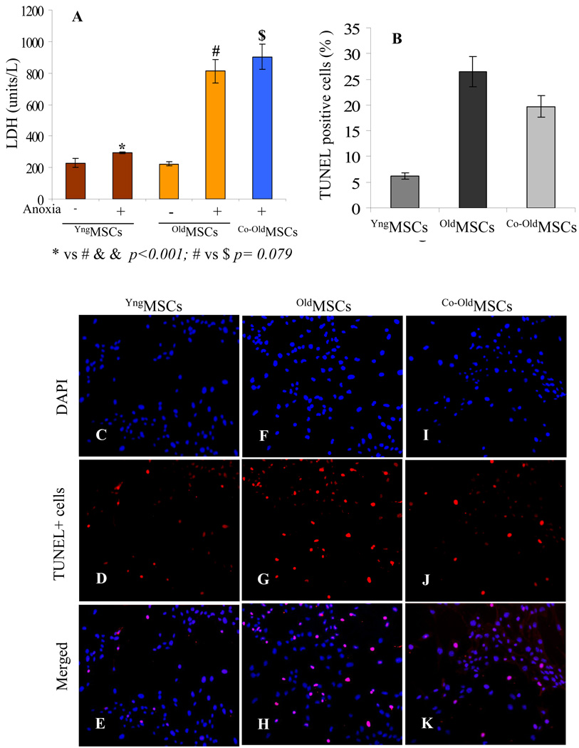
(A) LDH release from YngMSCs, oldMSCs and Co-oldMSCs as an indicator of cellular injury. YngMSCs were highly resistant to 8h anoxia as was indicated by the significantly low level LDH release as compared with the oldMSCs. oldMSCs in the presence of their younger counterparts (Co-oldMSCs) failed to show improved resistance to anoxia. YngMSCs and oldMSCs without exposure to anoxia were used as controls. (B–K) TUNEL assay on YngMSCs, oldMSCs and Co-oldMSCs after exposure to 8h anoxia showed that the number of TUNEL positive cells was significantly less in YngMSCs (C–E) as compared to oldMSCs (F–H) and Co-oldMSCs (I–K).

