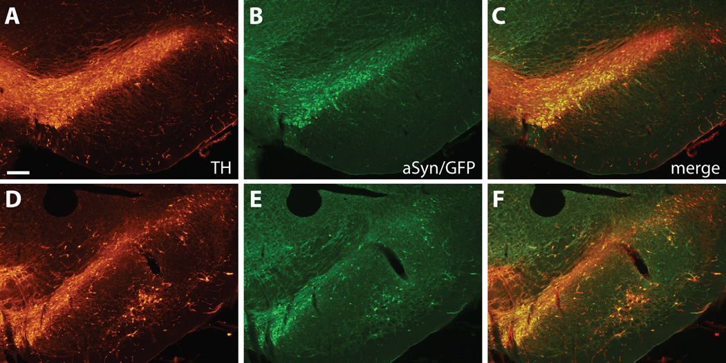
Figure 1.
Immunofluorescence staining of substantia nigra (SN) injected with rAAV2/8 expressing wt α-synuclein and internal ribosomal entry site-enhanced green fluorescent protein (EGFP). Tyrosine hydroxylase (TH) staining shows the distribution of dopaminergic SN neurons at representative rostral (A–C) and caudal (D–F) levels. GFP-positive transduced cells are widely distributed throughout the SN and almost exclusively colocalize with TH-positive neurons. Bar: 250 µm.

