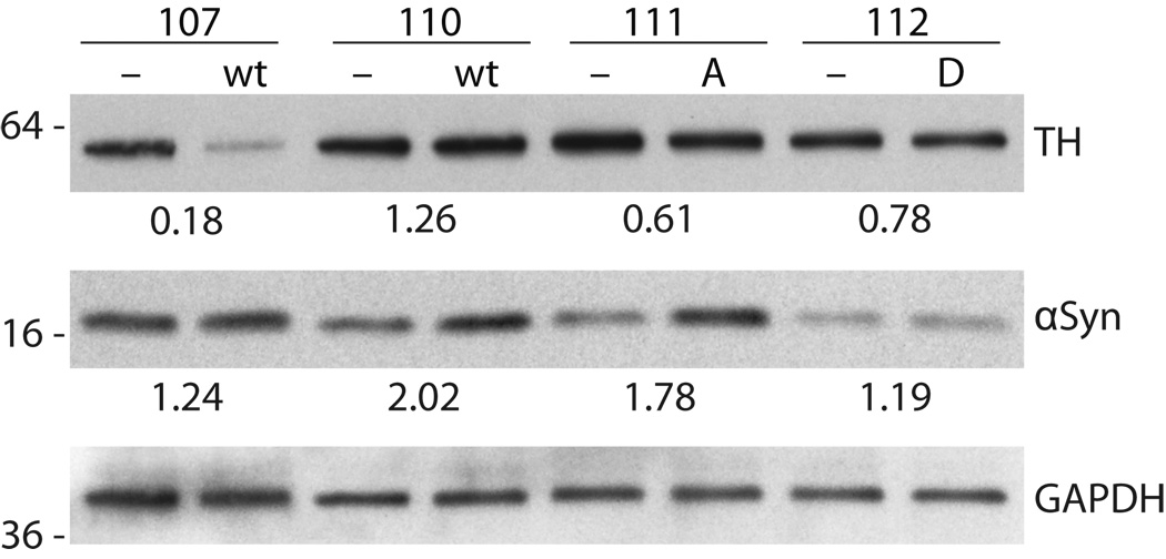
Figure 3.
Measurement of 〈-synuclein and tyrosine hydroxylase (TH) expression in midbrain tissue 6 weeks post-viral injection. Western blots show samples from midbrain ipsilateral to 〈-synuclein (wild type [wt], S129A [A], and S129D [D]) injection and are compared to protein extracts from the contralateral midbrain injected with empty vector (−) control in the same animal. Protein from left and right midbrain samples was probed with primary antibody to 〈-synuclein and TH, followed by secondary HRP-conjugated antibody and ECL detection. Bands were quantified and normalized to GAPDH loading control. The ratio of 〈-synuclein and TH for the α-synuclein (ipsilateral) versus empty vector (contralateral) side was calculated for each animal. Blots show 〈-synuclein overexpression for most animals, but there was variable expression as shown for the 2 wt samples. Decreased TH expression is also shown for wt and S129 mutants.

