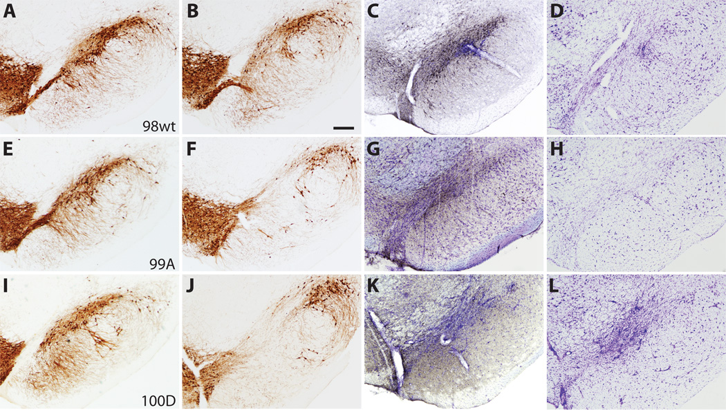
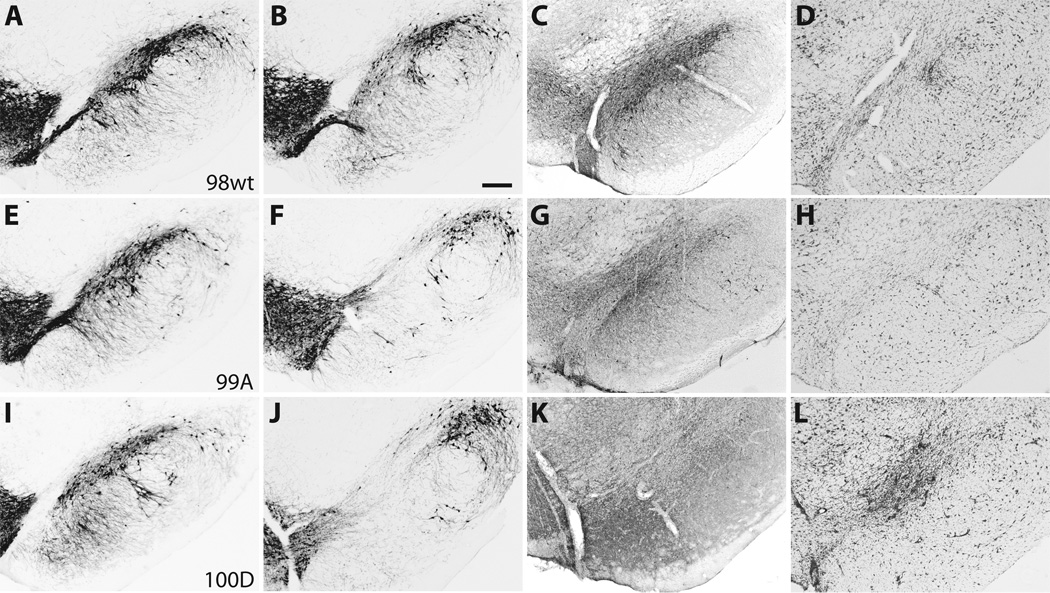
Figure 4.
Photomicrographs of representative substantia nigra (SN) sections at 6 weeks post-injection demonstrate neurodegenerative changes and tyrosine hydroxylase (TH) cell loss. (A–D) Case 98, wild type (wt) 〈-synuclein. (E–H) Case 99, S129A. (I–L) Case 100, S129D. (A, E, I) TH immunostaining in the contralateral SN after empty vector control injection. (B, F, J) TH cell loss in the SN for wt and S129 mutant cases. (C, G, K) Distribution of human 〈-synuclein (LB509) expression (gray-black immunostaining) within the SN. (D, H, L) Adjacent sections stained with cresyl violet that demonstrate SN cell loss and gliosis at rAAV injection sites. Bar: 250 ⌠m.


