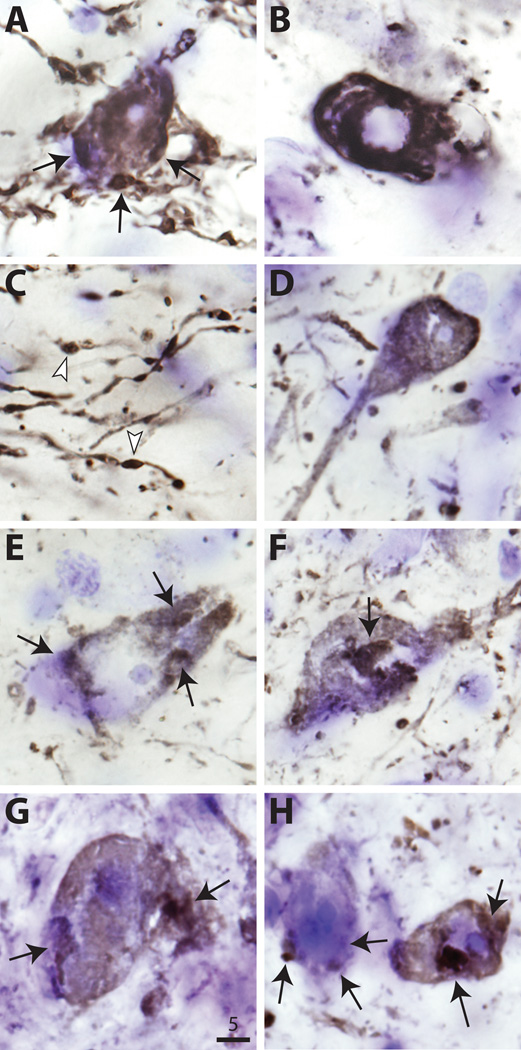
Figure 6.
High-power photomicrographs of human α-synuclein (LB509) immunostaining of nigral neurons in wild type (wt) (A–C), S129A (D–F), and S129D (G, H) recipients. Arrows indicate large intracytoplasmic 〈-synuclein-positive aggregates. Most aggregates were either perinuclear or in the periphery (A and G). B shows typical nuclear ghosting, consistent with neurodegenerative change. Dystrophic neurites in C have characteristic α-synuclein-positive inclusions (arrowheads). Bar: 5 ⌠m.

