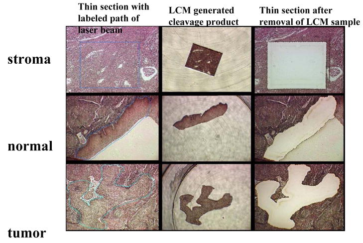
Fig. 1. Laser Capture Microdissection of one cluster each of stromal, normal squamous and cancerous cells from a thin section of a biopsy of a cervical squamous carcinoma.
The first column of three microscopic pictures shows the fixed tissue after thin section, a blue line indicating the programmed path of the laser beam. The second column shows the cleaved sample after transfer to a test tube. The third column the remaining tissue after removal of the experimental sample. The three cuts were targeted at the same thin section of a tumor.

