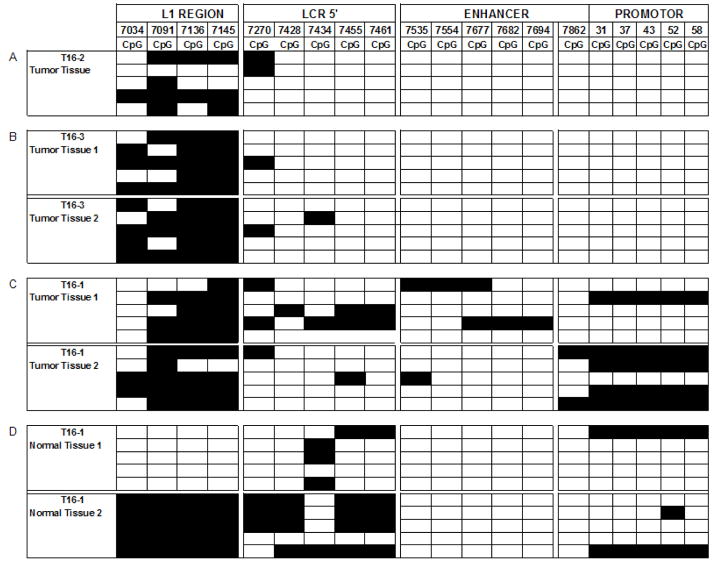
Fig. 2. Distribution of methylated CpG residues at the 3′ flank of L1 and through the LCR of HPV-16.
Each vertical set of rectangles represents one of 19 specific CpG dinucleotides, the number on the top of the bar the position of this CpG in the genome of HPV-16. Each horizontal set of rectangles represents a 913 bp segment of the HPV-16 genome, covering the 3′ end of the L1 gene and the complete long control region. Unmethylated CpGs are indicated by white rectangles, methylated by black ones. The two vertical white separators indicate the borders between amplicons, and discontinuities between supposedly different HPV-16 molecules. Further experimental details of this approach and interpretation of the preferential methylation of the L1 gene has been addressed in detailed by multiple studies from our group that targeted cervical, oral, and penile HPV-16 infections (Kalantari et al., 2004; Balderas-Loaeza et al., 2007; Kalantari et al., 2008).

