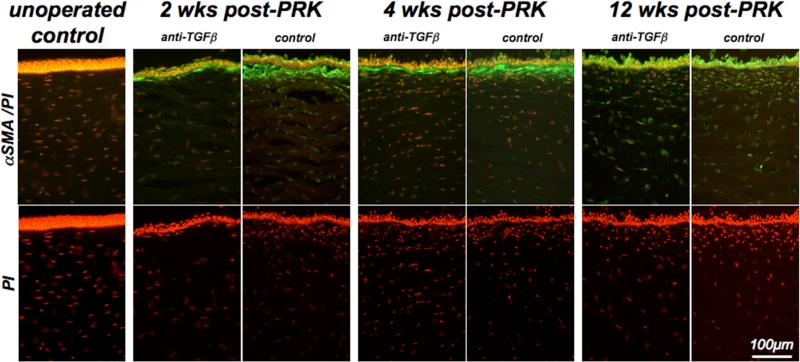
Figure 5. Effect of anti-TGFβ treatment on αSMA expression and stromal cell density.
Photomicrographs of corneal sections from a normal, unoperated cat and pairs of cat eyes that underwent PRK and received either vehicle or anti-TGFβ treatment post-operatively. These cats were sacrificed at 2, 4 and 12 weeks post-PRK and sections of their corneas were double-labeled with antibodies against αSMA to label myofibroblasts and with propidium iodide (PI) to label cell nuclei. Note the absence of αSMA staining in the operated cat cornea, in contrast with the significant αSMA expression in the sub-ablation stroma of eyes that underwent PRK. The band of αSMA expression was significantly thicker and more continuous in control eyes than in contralateral eyes treated with anti-TGFβ. It was also most intense at 2 and 4 weeks post-PRK, becoming almost absent in the stroma by 12 weeks-post-PRK. PI staining also revealed an area of increased cellularity under the ablation zone in all post-operative eyes, although cell density appeared consistently higher in control eyes relative to eyes treated with anti-TGFβ.

