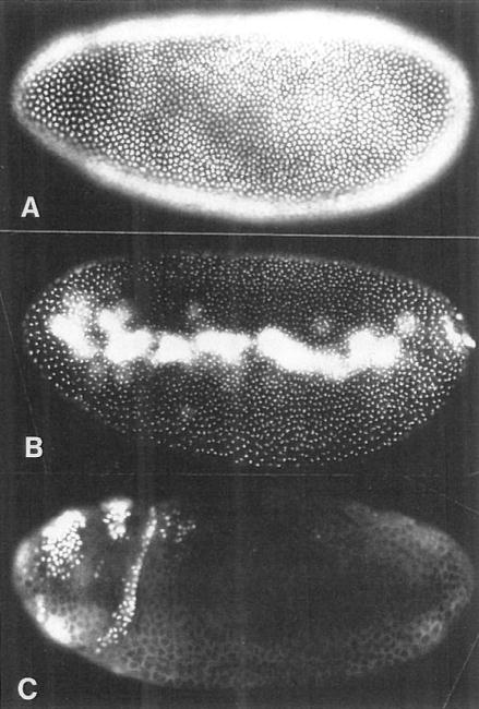
Figure 1. S Phase 14 Begins and Ends Synchronously without a G1 Period.
We show three embryos pulse labeled with BrdU for 20 min at progressively later stages of interphase 14. The BrdU is detected by immunoflourescence, and labeled nuclei appear white.
(A) An embryo labeled for the first 20 min of interphase 14 (130–150 min after egg deposition [AED]) shows all blastoderm nuclei heavily and uniformly labeled.
(B) An embryo labeled at about 160–180 min AED shows spots of late replicating centromeric heterochromatin in each blastoderm nucleus. Note that nuclei in all regions of the blastoderm are similarly labeled, indicating that S phase completion is not spatially patterned. The yolk nuclei and pole cell nuclei are heavily labeled in this embryo, indicating that they replicate their DNA later than the blastoderm nuclei.
(C) An embryo labeled at about 180–200 min AED shows most of the blastoderm nuclei unlabeled, indicating a true G2 period. Patches of labeled nuclei in the anterior are those that have already progressed through mitosis 14 and entered S phase 15. This photo is overexposed to show cytoplasmic background staining.

