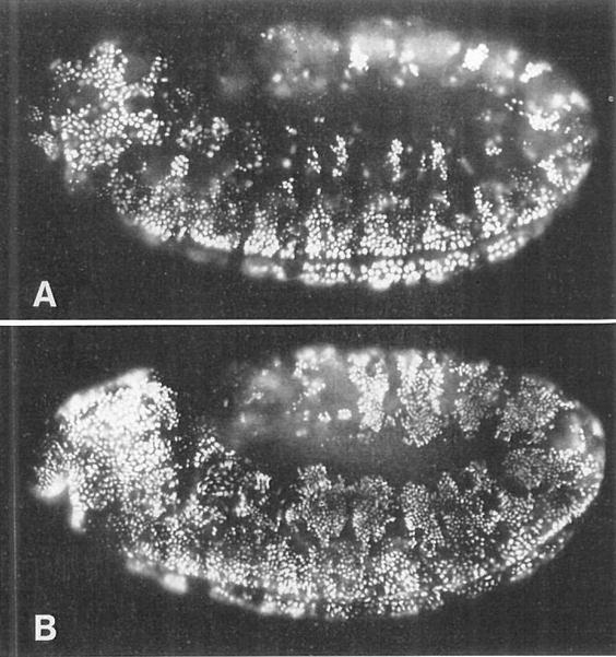
Figure 6. The Decision to Enter G1 of Cycle 17 Is Executed in G2 of Cycle 16.

(A) A wild-type embryo heat pulsed from 350–380 min AED and then labeled with BrdU for 30 min. This pattern is identical to that seen in wild-type embryos that were not heat pulsed. Ventral cells are completing S phase 16 and are heavily labeled. Dorsolateral cells have progressed through mitosis 16, but only a few of them (one small cluster per segment) have entered S phase 17 and become labeled. The majority of these dorsolateral cells do not label with BrdU after mitosis 16, but instead enter their first G1 period.
(B) A hs-stg embryo that was similarly heat pulsed and BrdU labeled. In this embryo, the dorsolateral cells have been induced to go through mitosis 16 prematurely, and many of them have become heavily labeled with BrdU. More than the normal number of labeled cells can also be seen in the head, to the left. Thus, premature entry into mitosis 16 triggers an unscheduled S phase 17. Mitotic and replication patterns in the ventral region are essentially unaffected. Both embryos are lateral views, with anterior to the left. These are germ-band extended embryos, so the dorsal-most tissue (the amnioserosa) is the region in the center of the embryo, and strips of dorsolateral tissue curl around it. Ventral tissues are at the periphery of the embryo, on both the top and the bottom.
