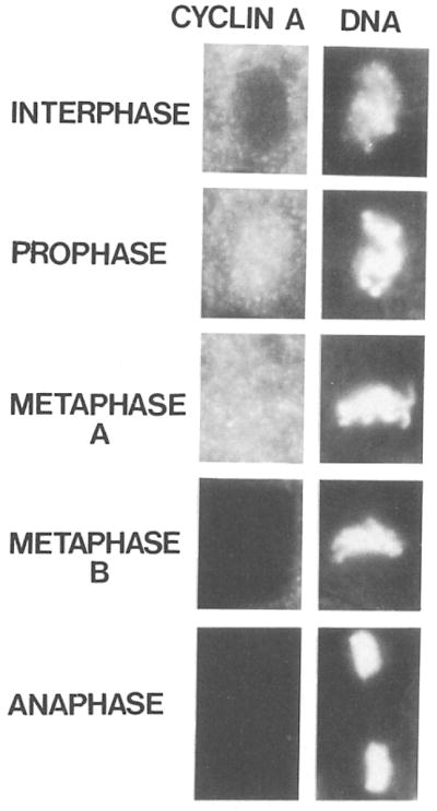
Figure 6. Intracellular Distribution of Cyclin A during Cell Division.

Embryos were double labeled with affinity purified antibodies against cyclin A followed by rhodamine conjugated secondary antibodies (left column) and with the DNA stain Hoechst 33258 (right column). Cells in interphase, prophase, metaphase, and anaphase are shown. Cyclin A is degraded within metaphase and therefore metaphases with cyclin A staining (Metaphase A) as well as without (Metaphase B) can be detected.
