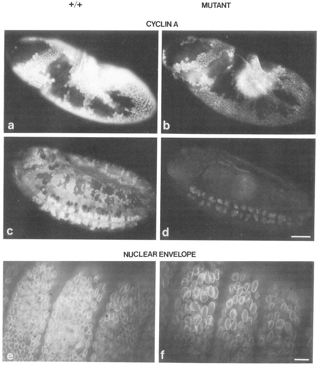
Figure 8. Cyclin A Distribution and Nuclear Density in Mutant Embryos.
Embryos were stained with affinity purified antibodies against cyclin A followed by rhodamine conjugated goat anti-rabbit antibodies (a–d) and with a monoclonal antibody against a nuclear envelope component followed by fluorescein conjugated goat anti-mouse antibodies (e and f). (a, c, and e) Normal embryos, (b, d, and f) Mutant embryos. Embryos are at the time of mitosis 14 (a and b), mitosis 15 (c and d), or after mitosis 16 (9 hr, e and f). The level of cyclin A staining in the progeny of the cross neo114/TM3 × vin3/TM3 is wild type (a and c) in 25%, intermediate (data not shown) in 50%, and clearly lower (b and d) in 25% of the embryos. Double labeling with anti–cyclin A antibodies (data not shown) and antinuclear envelope antibodies (e and f) revealed that embryos, which have no detectable cyclin A (f), have less than half the number of nuclei than in embryos where cyclin A was readily detected (e). The bar in (d) corresponds to 50 μm. Only the dorsolateral epidermis of three embryonic segments is shown in (e) and (f). The bar in (f) corresponds to 5 μm.

