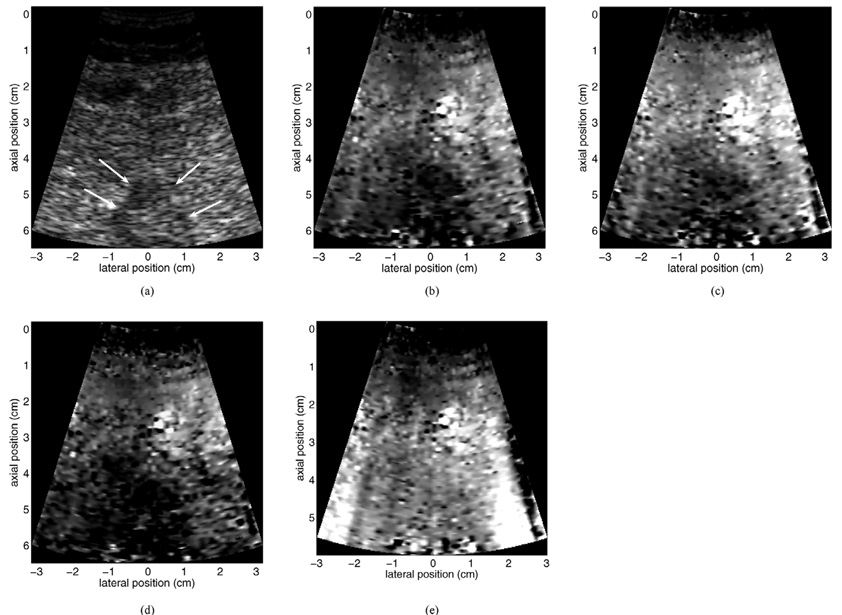
Fig. 13.
Motion filter comparison for data acquired from a 62-year-old female patient. Shown are B-mode (a) and linear RB motion filtered (b), MB motion filtered (c), and quadratic RB motion filtered (d) ARFI images of a thermal lesion created with RFA in the left hepatic lobe. The non-motion filtered raw displacement data are shown in (e) for reference. Note the significant motion artifacts in the non-motion filtered image. Arrows in (a) indicate boundaries of the thermal lesion in the B-mode image. All ARFI images are shown with the same displacement scale.

