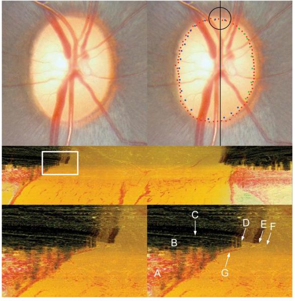
Figure 2. The identification of Bruch’s Membrane and Bruch’s Membrane Opening in a histomorphometric section.
Top left panel: The clinical disc photograph (OD) prior to disc margin delineation
Top right panel: The clinical disc photograph displaying both the co-localized histomorphometric BMO points (red glyphs) and the clinical disc margin points (blue and green glyphs). The black line is the approximate location of the vertical histomorphometric section shown in the middle panel. The area circled in black is the approximate location of the histomorphometric region (white box in the middle panel) magnified in the two bottom panels.
Middle panel: Central vertical histomorphometric section shown as a black line in the top right panel. Superior is left and inferior is right. The area within the white box is magnified in the bottom panels. Note that because the tissues are sectioned from the vitreous (top) to the orbital optic nerve (bottom) a dark shadow is present until the serial sectioning plane passes through the dense pigment of the retinal pigment epithelium, choroid and Bruch’s Membrane
Bottom left panel: Magnified view of the highlighted white box in the middle panel which demonstrates the superior disc margin anatomy
- A = Sclera
- B = Choroid
- C = Bruch’s Membrane
- D = Commencement of pigmented Bruch’s Membrane which in this section appears to co-localize to the termination of the choroid (B)
- E = Termination of pigmented Bruch’s Membrane and the commencement of unpigmented Bruch’s Membrane. Note the presence of pigment ‘shadows’ of variable density cast vertically along the course of the pigmented Bruch’s Membrane which are absent in the regions where Bruch’s Membrane is unpigmented
- F = Termination of unpigmented Bruch’s Membrane, which would be delineated as Bruch’s Membrane Opening in this section
- G = Border Tissue of Elschnig. In this eye, Bruch’s Membrane fuses with the superior edge of the Border Tissue and extends slightly beyond its termination

