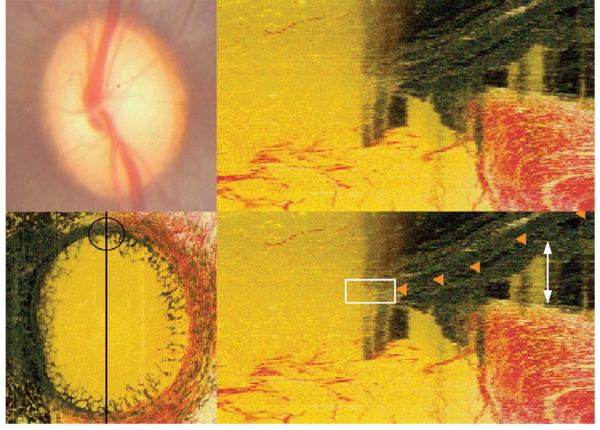
Figure 3. Pigment on the lamina surface causing obfuscation of Bruch’s Membrane Opening.
Top left panel: Clinical disc photograph (OS)
Bottom left panel: En face view of the histomorphometric reconstruction of the same eye. Black line shows the orientation of a histomorphometric section image, a portion of which (the superior disc margin) is magnified in the right panels. The black circle highlights the region viewed in the right panels; note the presence of pigment on the lamina surface
Top right panel: Histomorphometric view of the superior part of the neural canal
Bottom right panel: Bruch’s Membrane delineated (orange glyphs). The white rectangle highlights an area where the view of Bruch’s Membrane is obscured by a dark shadow cast from the lamina pigment below. In this circumstance, accurate delineation of Bruch’s Membrane Opening can be difficult. The white arrowheads highlight the extent of an artifactual choroidal detachment, most likely caused by the perfusion fixation process

