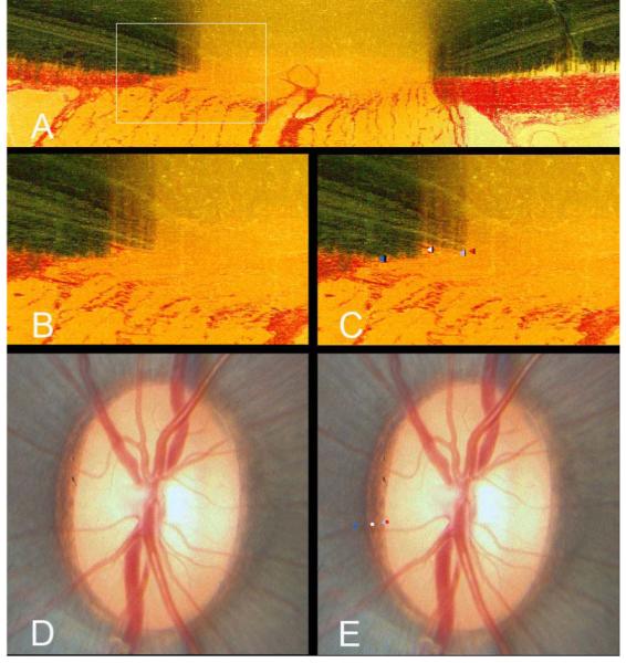
A histomorphometric section taken at 67.5° from a left eye. A white box highlights the area of interest in the nasal region of the optic nerve head
The area within the white box has been magnified to highlight the structures comprising the disc margin. In this section, there is a substantial overhang of Bruch’s Membrane (both pigmented and unpigmented) beyond the termination of the Border Tissue of Elschnig
The structures comprising the disc margin have been delineated within Multiview; termination of unpigmented Bruch’s Membrane, in other words Bruch’s Membrane Opening (red glyph), termination of pigmented Bruch’s Membrane (light blue glyph), junction of Border Tissue of Elschnig with Bruch’s Membrane (scleral ring of Elschnig - white glyph) and the anterior scleral canal opening (dark blue glyph)
The in vivo disc photograph of the eye from which the histomorphometric reconstruction was obtained
-
Following co-localization of the three-dimensional vessel reconstruction to the disc photograph, the termination of unpigmented Bruch’s Membrane (red glyph) coincides with the innermost white reflective halo at the disc margin. The termination of pigmented Bruch’s Membrane (light blue glyph) coincides with the inner edge of the pigment at the disc margin. The Border Tissue/Bruch’s Membrane junction (scleral ring - white glyph) coincides with a white reflective stripe within the mottled disc margin pigment; the anterior scleral canal opening (dark blue glyph) is external to this.
It is apparent in this eye that the innermost white reflective structure at the disc margin is not the scleral ring of Elschnig but the edge of unpigmented Bruch’s Membrane. In this region of the disc, the scleral ring is considerably external to what the clinician perceives as the disc margin

