
Figure 1.
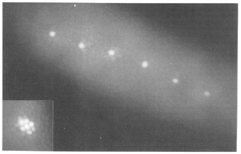
Single Nuclei Marked by Photoactivation
Live cellular blastoderm viewed in epifluorescence microscopy. The embryo was injected with nonfluorescent Dx-CF-NLS before cellularization. Single nuclei were made fluorescent during cellularization by illumination with a microbeam of 365 nm light. Inset: to show nuclear localization of the activated fluorescence, a group of nuclei in a different embryo were marked with a wider microbeam.