
Fig. 3. Lgl phosphorylation can regulate oocyte polarity.
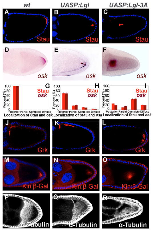
Stau (A-C), osk (D-F), the quantification of Stau and osk mRNA localization (G-I), Grk (J-L), Kin:β-Gal (M-O), α-Tubulin (P-R) in the wild type (driver only) (A,D,G,J,M,P), Lgl-overexpressing (B,E,H,K,N,Q) and Lgl-3A-overexpressing (C,F,I,L,O,R) egg chambers are shown. In egg chambers with Lgl overexpression, Stau (B,H), osk (E,H) and Kin:β-gal (N) were partially mislocalized, but Grk (K) showed normal localization, and α-Tubulin (Q) displayed a weaker anterior-posterior gradient than in the wild type (P). In egg chambers with Lgl-3A overexpression, Stau (C,I), osk (F,I) and Kin:β-gal (O) were frequently mislocalized to the center of the oocyte, Grk and the oocyte nucleus (L) were mislocalized in some egg chambers, and α-Tubulin showed higher concentrations along the cortex and lower concentration at the center of the oocyte (R).