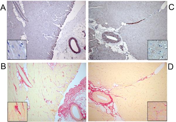
Figure 6. CML content in Left Ventricle (LV).
CML (A) and picros-sirius (B) staining of sequential LV sections (original10x) in a representative young normal (YN) dog showing lack of CML staining in myocardium or myocardial or peri-vascular collagen. Insert in A-D show higher magnification of myocardium. There is staining in vascular smooth muscle cells. CML (C) and picros-sirius (D) staining of sequential LV sections (10x) in a representative elderly hypertensive (OH) dog showing lack of CML staining in myocardium or myocardial or peri-vascular collagen. There is staining in vascular smooth muscle cells.

