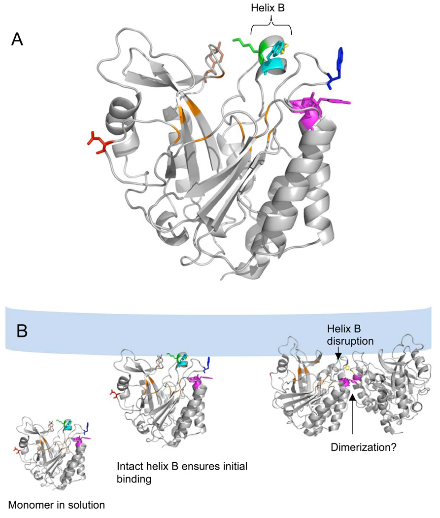
Figure 1.
(A) B. thuringiensis PI-PLC monomer structure (from the Y247S/Y251S crystal structure (14)) highlighting the active site pocket in orange and the surface residues altered in this work (Asn168, red; Pro42, yellow; Lys44, green; Tyr88, brown; Tyr246, Tyr247, Tyr248, magenta; Trp47, cyan and Trp242, blue). The short helix B contains Lys44 and Trp47 and is capped by Pro42 at the N-terminal end. (B) Proposed roles of the mutated residues in PI-PLC vesicle binding and activation (which may include dimerization). PI-PLC is a monomer in solution. After the initial Lys44 mediated attraction to the anionic membrane, an intact helix B orients Trp47 for membrane insertion. Subsequent disruption of helix B allows Pro42 to help stabilize the PI-PLC homodimer interface including a Tyr zipper involving Tyr residues 246–248. The dimer structure in this model is based on the W242A/W47A B. thuringiensis dimer crystal structure (12). The lipid bilayer is shown schematically in light blue.

