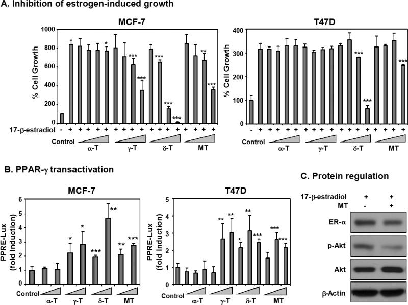
Fig. 4.
Mixed tocopherols (MT), γ-tocopherol (γ-T) and δ-tocopherol (δ-T), exert anti-estrogenic activity and activate PPAR-γ in ER-positive human breast cancer cells. A. Inhibition of estrogen-induced growth of MCF-7 and T47D by mixed tocopherols and individual tocopherol isoforms. MCF-7 and T47D cells were treated with tocopherols (10, 30, 60, and 100 μM) in 10% charcoal stripped FBS/phenol red free RPMI medium for 3 days, together with 17-β-estradiol (10 pM), and the cell proliferation was compared using [3H]thymidine uptake assay. Statistical significance, *p < 0.05, **p < 0.01, ***p<0.001. B. PPAR-γ transactivation by mixed tocopherols and individual tocopherol isoforms. MCF-7 and T47D cells were transfected with DNA vectors (50 ng PPAR-γ, 40 ng PPRE-Luc, 50 ng, RXR-α, and 10 ng β-gal in 24-well plates) for 4 h in serum-free media. Then, cells were treated with compounds (50 and 100 μM in MCF-7 cells and 20, 50, and 100 μM in T47D cells) for additional 36 h in 10% charcoal stripped FBS in phenol red free RPMI. Luciferase activity was measured with a luminometer and normalized by β-galactosidase activity. Statistical significance, *p < 0.05, **p < 0.01, ***p<0.001. C. The effect of protein expression by mixed tocopherols (100 μM) in the presence of 17-β-estradiol (10 pM) in MCF-7 cells. After the incubation of 17-β-estradiol and mixed tocopherols for 24 h, the protein levels of ER-α, p-Akt, Akt, and β-actin were determined.

