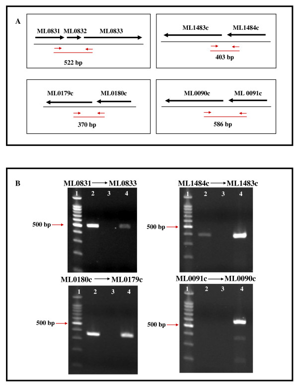
Figure 3.
Read-through transcription of M. leprae pseudogenes. This figure represents the results of RT-PCR analysis of transcriptional read-through between M. leprae pseudogenes and their upstream ORFs. Panel A shows mapped genomic regions where pseudogenes (ML0832, ML1483c, ML0179c, and ML0091c) and upstream ORFs are located http://genolist.pasteur.fr/Leproma/. Black arrows indicate direction of transcription. Small red arrows indicate forward and reverse primers for RT-PCR. Red line below primers and (bp) designate size of predicted PCR product if genes are expressed as a polycistronic mRNA. Panel B shows ethidium bromide-stained agarose gel analysis of these PCR amplicons: Lane 1, 100 bp DNA ladder (New England Biolabs); Lane 2, PCR amplicons from nu/nu footpad-derived M. leprae Thai-53 cDNA for each gene set; Lane 3, RT (-) control; Lane 4, 1 ng M. leprae DNA (positive control).

