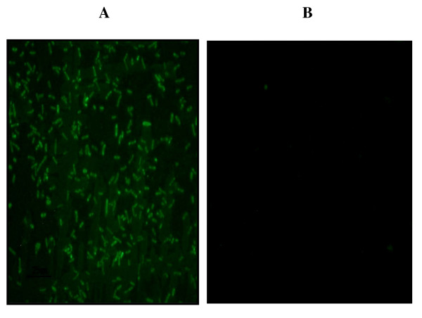
Figure 5.
In vitro promoter activity in pyrR (ML0351) pseudogene. Panel A shows an image of GFP-fluorescent E. coli::pGlow-TOPO-TA/MLpyrR prom as a result of transformation of E. coli XL-1 Blue cells with the pGlow-TOPO-TA (promoterless and lacking a SD site) containing the upstream region of the M. leprae pyrR pseudogene; including the promoter, SD and start codon (Additional File 6, Fig. 3A) fused into the ATG of gfp. A clone containing the ampicillin-resistant phenotype and positive for the M. leprae pyrR promoter insert by PCR/DNA sequencing was analyzed by fluorescent microscopy using a Nikon Eclipse E400 fluorescent microscope using a GFP filter (excitation/emission maxima of 480 nm/560 nm). Panel B shows an image of E. coli::pGlow-TOPO-TA re-circularized vector (negative control for background fluorescence).

