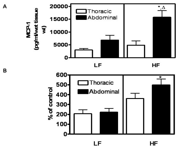
Figure 3. MCP-1 release (A) and macrophage migration (B) in periaortic adipose tissue explants from abdominal aortas are increased by obesity.

Periaortic adipose tissue explants were prepared from thoracic or abdominal aortas of LF- and HF-fed mice (4 months) and incubated as described. A, MCP-1 release into the media was increased in periaortic explants of abdominal aortas from HF compared to LF-fed mice. There was no effect of HF-feeding on MCP-1 release from thoracic periaortic adipose tissue explants. B, Mouse peritoneal macrophage migration was increased by conditioned media from abdominal periaortic adipose explants of HF compared to LF-fed mice.*, significantly different from LF abdominal.  , significantly different from HF-thoracic. Data are mean ± SEM from n = 5 mice/group.
