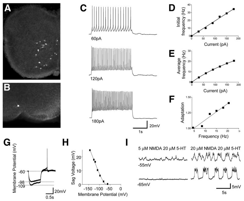
Figure 3. Cellular properties of V3-derived neurons in lamina VIII.
(A) Vibratome slice from P0 Sim1Cre; ZnG spinal cord showing V3-derived neurons expressing GFP.
(B) An example of a whole cell patch-clamp recorded V3 neuron labeled with neurobiotin.
(C and D) Representative response of a ventral V3-derived neuron to a series of 2s depolarizing currents (C). A linear relationship was found in the initial spike frequency as a function of the increasing input currents (D).
(E) The relationship of the average spike frequency as a function of the input current was fitted by a non-linear regression function (Y=a*x/(1+x/b)).
(F) A moderate but linear increase in the spike frequency adaptation was found along the increasing spike frequency (n==14 cells). The level of spike adaptation was determined by the average of the last three spike frequencies divided by the average of the first-three spike frequencies for varying 2 second current steps. Current steps of 20–210 pA were applied to each cell.
(G-H) Small to moderate sag voltages and post rebound potentials were produced by a series of 1 s hyperpolarizing currents in lamina VII/VIII V3 neurons. The amplitude of the sag voltage was strongly voltage dependent (H).
(I) Some V3-derived neurons display slow oscillations in membrane potential in the presence of 20 μM NMDA/20 μM 5-HT but not 5 μM NMDA/20 μM 5-HT.

