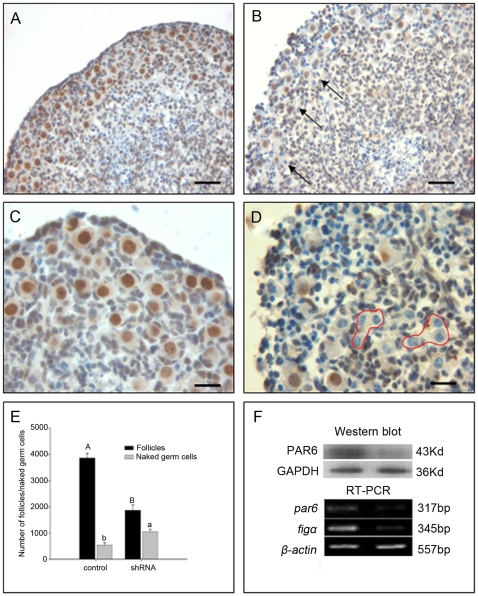
Figure 4. Loss of PAR6 disrupted ovarian histogenesis and reduced follicular formation.
15.5 dpc ovaries were treated with control shRNA plasmid or shRNA for 12 hours, cultured for 6.5 days, then the shape of ovaries and the localization of PAR6 (A and C, control; B and D, shRNA ),the number of follicles and naked germ cells (E), the expression of mRNA and protein (F) were examined. Arrows noted the naked germ cells and red rounds noted the cysts with PAR6 negative germ cells. Each data came from five ovaries, different superscripts denote differences (P<0.05) respectively. RT-PCR and Western blot were repeated at least three times with similar results. A and B, Bar = 40 µm; C and D, Bar = 20 µm.

