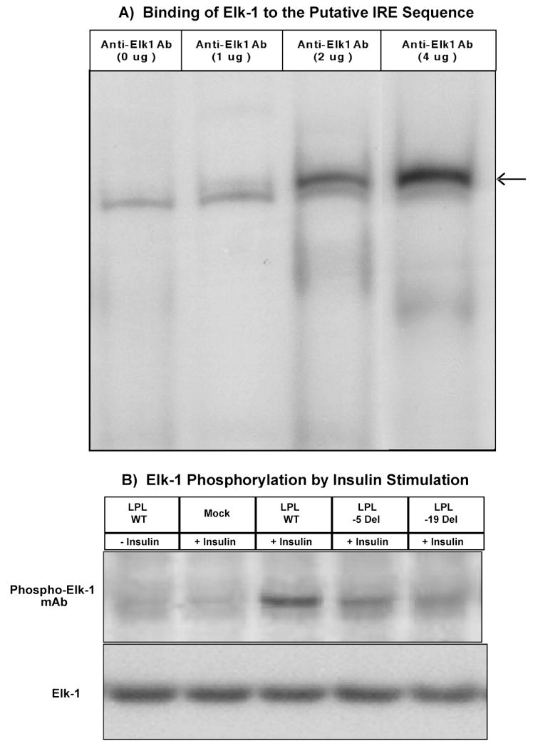
Fig. 5.

(A) Binding of Elk-1 to the Putative IRE Sequence. Super Gel Shift analysis. 29-bp DNA fragment from the putative IRE region in exon 10 of the LPL gene was used as 32P-labeled probe. Nuclear extracts prepared from human aorta smooth muscle cells were incubated in the absence (0 μg) or presence (1μg, 2 ug and 4 μg) of anti-Elk-1 antibody added prior to gel mobility shift analysis. Please note that in the absence of anti-Elk1 antibody (0 μg) there is only one band. However, in the presence of 1 ug of anti-Elk1 antibody a small supershift band appeared whose intensity increased proportionately in the presence of 2 μg and 4 μg of anti-Elk1 antibody, respectively (super shift band is indicated by an arrow). (B) Elk-1 Phosphorylation by Insulin Stimulation. Nuclear protein from human aorta smooth muscle cells transfected with expression plasmids for exon 10 wild type, 5 bp deletion and 19 bp deletion mutant, treated with carrier control (25 μM HEPES, − insulin) or insulin (+ insulin) for 30 min. and then subjected to Western blot analysis using Phospho-Elk-1 (Ser383) monoclonal antibody (upper panel).
