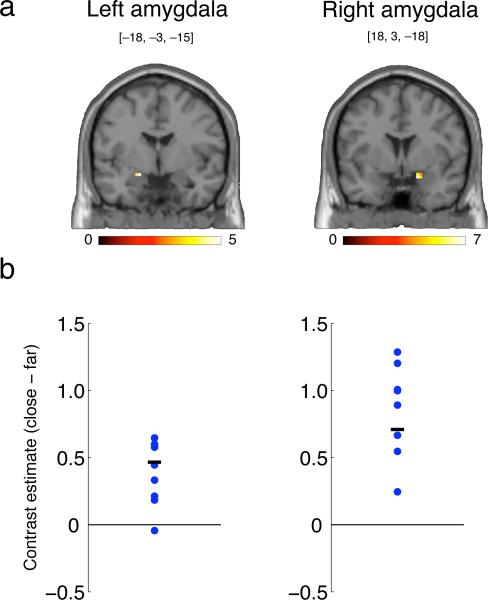
Figure 2.
fMRI study: Activation of the amygdala by close (relative to far) interpersonal distance. (A) Coronal slices showing significantly activated voxels in the dorsal amygdala (cluster-level significance, p<0.05); scale shows t-value. (B) Contrast parameters (arbitrary units) for each of the eight subjects who participated in the experiment (extracted from and averaged across all significant voxels in (A); blue dots), along with the group mean (black line). Coordinates for the peak voxel are shown. Subjects were unable to see the position of the experimenter, but were informed of his location at all times. All experiments were approved by Caltech's Institutional Review Board, and informed written consent was obtained from all participants. See Supplementary Text for a detailed description of the experiment.

