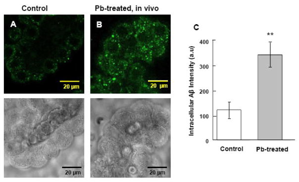
Fig. 1.
Increased accumulation of intracellular Aβ in rat choroid plexus tissues following in vivo acute Pb exposure by confocal study. (A). Choroid plexus tissue from a control rat. (B). Rats received a single ip injection of 27 mg Pb/kg. Twenty-four hr post exposure, FAM-labeled Aβ1–40 was infused into brain ventricles for 0.5 min. The plexus tissues were then removed 20 min post infusion for the confocal study. Substantial stains were evident in the cytosol but not in nuclei of choroidal epithelia. The lower panel shows the corresponding transmission image, indicating normal morphology of plexus tissues. (C). Quantification of the fluorescent signals using Laser Scanning Cytometry. Data represent mean ± SD, n=16 (a total of 16 cells per group taken from 4 tissue samples with fluorescence averaged from 4 cells per sample). **: p<0.001 as compared to controls.

