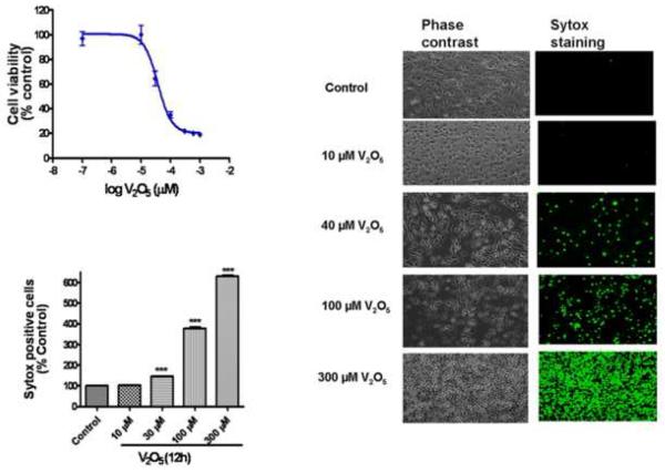
Fig. 1.
Vanadium induces a dose-dependent neurotoxic effect on N27 dopaminergic neuronal cells. (A) Effect of vanadium on cell viability in N27 dopaminergic neuronal cells. The cells were exposed to 0-300 μM V2O5 and then cell viability was measured using the MTT assay. The value of vanadium neurotoxicity in N27 dopaminergic cells was EC50 = 37 ± 3.47 μM. Data represent results from at least eight individual measurements. (B) Visualization of vanadium-induced neurotoxicity by Sytox green fluorescence assay. N27 dopaminergic neuronal cells were exposed to 40 μM V2O5 for 12 h and then cells were loaded with Sytox green and observed under a Nikon inverted fluorescence microscope and pictures were captured with a SPOT digital camera (Diagnostic Instruments, Sterling Heights, MI). (C) Quantitative analysis of vanadium-induced neurotoxicity was measured by the Sytox green cytotoxicity fluorescence assay. The Sytox fluorescence was measured by Bio-Tek fluorescence microplate reader. Data represent results from at least eight individual measurements and are expressed as mean ± S.E.M. The values are expressed as a percentage of untreated control cells. **p<0.001 indicates significant difference with each of the other groups.

