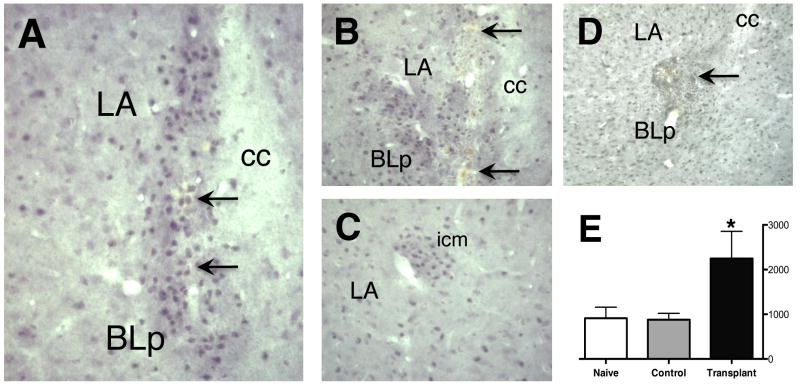
Figure 3.
GABA immunoreactivity illustrates large numbers of engrafted GABAergic neurons within the LBA (A, B). Graft sites are confirmed by the presence of fluorescent microspheres (see Fig. 3) and by the presence of blood by-products from the injection process (arrows). C, grafts are easily distinguished from endogenous intercalated cell masses (icm), which are more organized, typically comprised of fewer cells, and are devoid of fluorescent microspheres and evidence of trauma from the delivery cannula. D, a control graft of freeze-thaw killed LGE suspension shows disruption and scarring of the BLC and few GABA-ir neurons at the injection site. E, Bioquant analysis demonstrated a significant increase in overall GABA neuron density within the LBA (cells/mm2) for Transplant animals (*p < 0.05).

