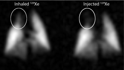
Figure 5:
Ventilation (left) and injected (right) 129Xe MR images acquired with an identical sequence and imaging parameters (1 × 1-mm resolution). This particular rat showed reduced ventilation in the right cranial lobe of the lung and matching signal intensity reduction on the injected 129Xe image (circled areas); this likely resulted from reduced perfusion in the same area.

