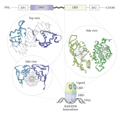
Figure 2.
Domain organization and structural binding modes of NRs. Upper panel: NRs are composed of an N-terminal regulatory domain (activation function 1 = AF1), followed by a DNA-binding domain (DBD), a ligand-binding domain (LBD), and a C-terminal domain (activation function 2 = AF2). Left panel: 3D model illustrating how the DBDs of the RAR/RXR heterodimer (PDB 1DSZ) interact with their target DNA-sequence. Right panel: solid ribbon representation illustrating the LBD of the RAR/RXR heterodimer (PDB 1DKF) complexed with the ligands 9-cis-RA for RXR (PDB 3LBD) and ATRA for RAR (PDB 2LBD). PDB files are taken from the RCBS Protein Data Bank (http://www.pdb.org/).

