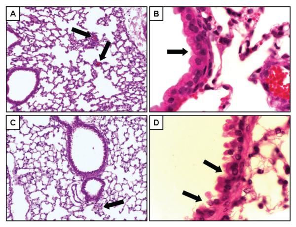
FIG. 4.

Assessment of lung histopathology following acute exposure to vaporized MA. Lung morphology was evaluated by H&E staining of paraffin sections. Representative sections from control mice (n = 4; panels A—B) and mice 3 h after MA exposure (n = 4; panels C—D). Panels A and B show representative images of lung tissue (original magnification, 20×). In panel A, the capillaries (black arrows) are apparent throughout the tissue. In panel C, no clear capillaries could be identified, but what appears to be the remains of a small arteriole is shown (black arrow). Higher magnification (original magnification 100×) of the airway epithelial cells are shown in panels B and D. The black arrows in panel D point to areas of disruption of normal cellular architecture.
