
Fig. 11.
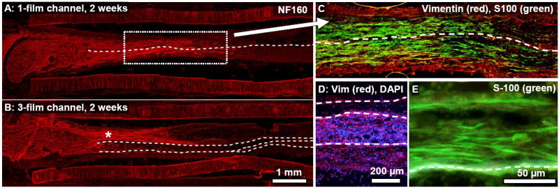
Longitudinal sections from a 1-film (A,C) and 3-film channel (B,D,E), two weeks post-surgery. (A,B): NF160+ axons had regenerated approximately one third of the distance through each channel type. Asymmetric growth of the regenerating axons into the topmost zone of this 3-film channel (‘*’ symbol) might have been due in part to the off-center location of the proximal nerve stump. Regeneration in the 1-film channel (A,C) was symmetrically distributed around the single thin film, although this was not always the case. (C): Magnified view of the boxed region in (A). Fibroblasts had fully bridged the gap. (D): Image from near the midpoint of the 3-film channels. Fibroblasts had fully bridged the gap and were distributed asymmetrically between the different zones of the 3-film channel (image taken near channel midpoint.) (E): Cross-section taken towards the distal end of the 3-film channel. Schwann cells, seen here migrating from the distal end, were highly aligned along the aligned thin-films, growing in direct contact with the thin-films, on top of each other, and also through the tissue matrix of the developing regeneration cable.