
Fig. 12.
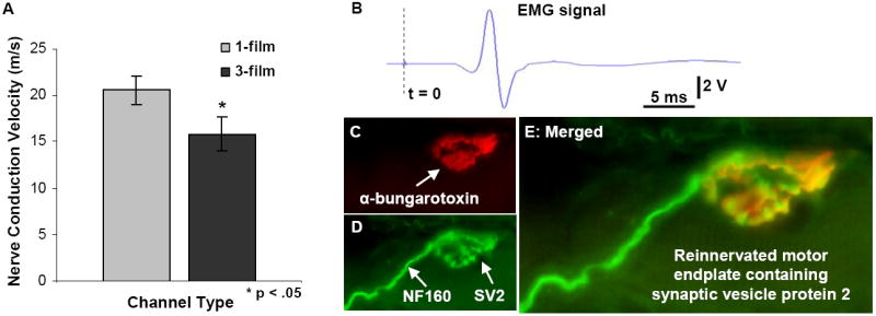
Evaluation of nerve conduction velocity and muscular reinnervation. (A): Significantly higher compound action potential conduction velocities were measured from nerves regenerated through the 1-film channels. (B): All gastrocnemius muscles produced measurable EMG signals and visible contractions. (This representative trace was taken from a 3-film channel case.) (C-E): As a further evaluation of muscular reinnervation, sectioned gastrocnemius muscles were reacted for immunofluorescent demonstration of (C) motor endplates, (D) axons, and vesicles containing synaptic vesicles 2 (SV2) protein. (E) Co-localization of these three markers confirmed the presence of reinnervated motor endplates in all treated animals.